01
Small Bowel Tumours
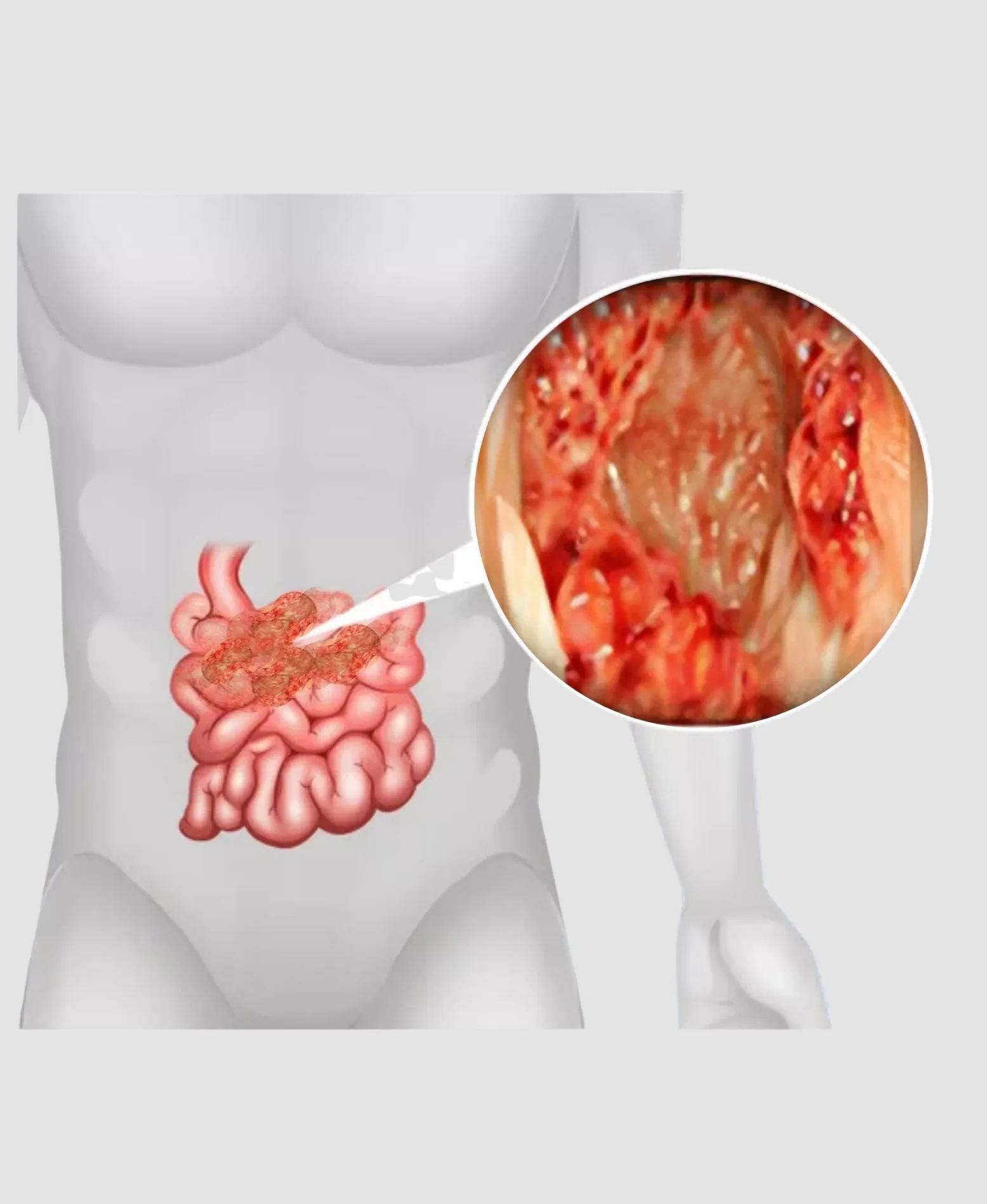
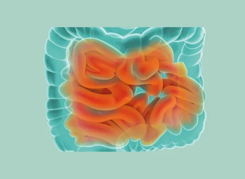
Tumours of the small intestine are rare - accounting for fewer than 5% of all gastrointestinal tumours - but are important because they are often diagnosed late (due to the relative inaccessibility of the small bowel and the non-specific nature of their symptoms) and because a significant proportion are malignant.
Types of small bowel tumours:
Carcinoid Tumours (Neuroendocrine Tumours - NETs):
The most common small bowel malignancy. Carcinoids arise from enterochromaffin cells (neuroendocrine cells) in the intestinal mucosa - most commonly in the terminal ileum (the last segment of the small intestine, joining the large intestine at the ileocaecal valve). Many carcinoid tumours are small and slow-growing but can metastasise to the liver and lymph nodes. When metastatic, they may produce the carcinoid syndrome - episodic flushing (redness of face and neck), diarrhoea, wheezing, and right heart disease - from secretion of serotonin and other vasoactive amines directly into the systemic circulation.
Surgical resection of the primary tumour with regional lymph node dissection is the treatment of choice. Even in the presence of hepatic metastases, debulking surgery (cytoreduction) may provide significant symptomatic relief and prolong survival.
Gastrointestinal Stromal Tumours (GISTs):
GISTs are mesenchymal tumours arising from the interstitial cells of Cajal (the pacemaker cells of the intestinal wall). In the small bowel, GISTs occur most commonly in the jejunum and ileum and present with occult or overt bleeding, abdominal pain, or a palpable mass. Risk stratification (based on size, mitotic rate, and location) guides treatment: small, low-risk GISTs may be observed; larger or higher-risk tumours require laparoscopic wedge resection (removing the segment of bowel bearing the tumour with clear margins). Imatinib (a tyrosine kinase inhibitor targeting the KIT mutation present in most GISTs) is used as adjuvant therapy for high-risk tumours.
Adenocarcinoma of the Small Bowel:
The second most common small bowel malignancy. Most frequently affects the duodenum (particularly the periampullary region) and the proximal jejunum. Presents with obstructive jaundice (periampullary), abdominal pain, weight loss, occult bleeding, and intestinal obstruction. Surgical resection (pancreaticoduodenectomy for duodenal adenocarcinoma; segmental small bowel resection for jejunal and ileal tumours) is the treatment of choice for resectable disease. Prognosis depends on stage and margin status.
Lymphoma of the Small Bowel:
Primary gastrointestinal lymphomas - predominantly MALT lymphoma (Mucosa-Associated Lymphoid Tissue, linked to H. pylori or celiac disease) and diffuse large B-cell lymphoma - can involve the small intestine. Presents with abdominal pain, diarrhoea, malabsorption, and occasionally perforation or obstruction. Diagnosis requires full-thickness biopsy. Management is primarily chemotherapy-based, with surgery reserved for complications (perforation, bleeding, obstruction).
Benign Small Bowel Tumours:
Lipomas, haemangiomas, adenomas, and inflammatory fibroid polyps are rare but may cause intussusception (telescoping of a bowel segment into the adjacent bowel - the tumour acts as a "lead point"), obstruction, or bleeding. Laparoscopic segmental resection is performed for symptomatic tumours.
Treatment Approach
Investigation of suspected small bowel tumours:
The small bowel's length and inaccessibility make conventional endoscopy inadequate for visualisation beyond the duodenum and terminal ileum. Key investigations include:
- CT enterography (CTE): CT with oral contrast to distend the small bowel - the best cross-sectional imaging modality for small bowel tumours and masses
- MR enterography (MRE): MRI-based alternative without radiation - preferred for younger patients and for repeated monitoring
- Capsule endoscopy: The patient swallows a pill-sized wireless camera that photographs the entire small bowel mucosa as it travels through - the best modality for identifying mucosal sources of obscure gastrointestinal bleeding
- Balloon-assisted enteroscopy (single or double balloon): Allows both visualisation and biopsy/treatment of small bowel lesions using a specialised endoscope advanced progressively through the small bowel
- PET-CT and octreotide scan (Somatostatin receptor scintigraphy): For staging and identifying metastatic sites in carcinoid tumours