
01
Pancreatic Cyst
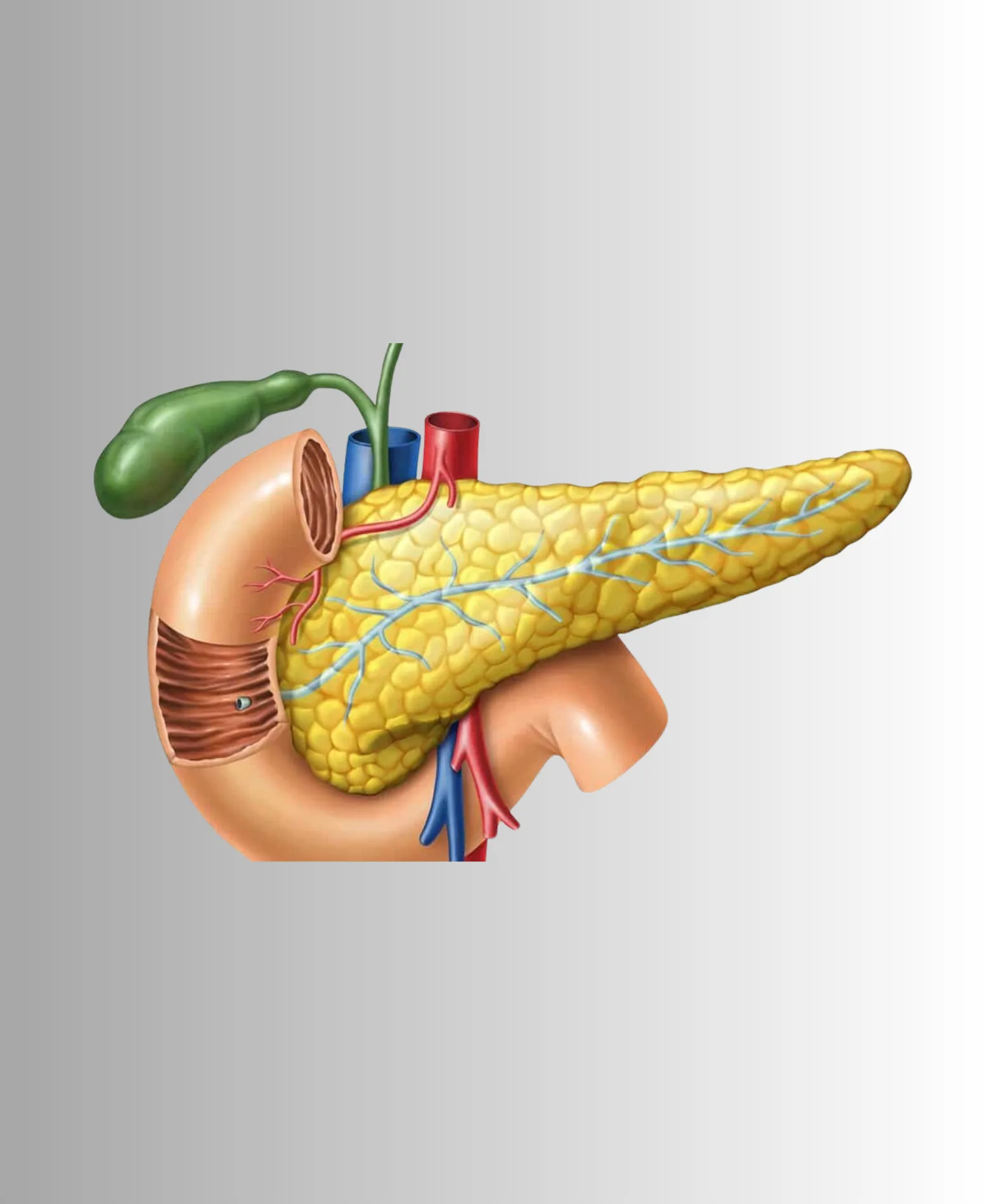
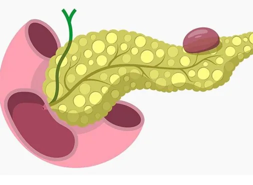
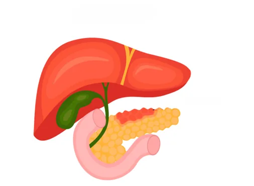
Pancreatic cysts are fluid-filled lesions within or adjacent to the pancreas - discovered increasingly often as abdominal imaging is performed more frequently. They range from entirely benign pseudocysts (accumulated fluid from pancreatitis) to cystic pancreatic tumours with significant malignant potential. Accurate differentiation is critical to appropriate management.
Pancreatic Pseudocyst:
The most common pancreatic cystic lesion - a collection of pancreatic juice and debris encapsulated by a fibrous wall of granulation tissue, forming as a complication of acute pancreatitis (fluid from a disrupted pancreatic duct becomes walled off) or trauma to the pancreas. Pseudocysts lack a true epithelial lining (hence "pseudo" cyst).
Most pseudocysts resolve spontaneously over 4-6 weeks without intervention. Indications for drainage include: failure to resolve, enlargement, causing symptoms (pain, early satiety, gastric outlet obstruction, jaundice from bile duct compression), or infection (abscess formation). Drainage is performed endoscopically (endoscopic transmural drainage - creating a connection between the pseudocyst and the stomach or duodenum using an endoscope) or laparoscopically (laparoscopic cystogastrostomy or cystojejunostomy).
Cystic Pancreatic Tumours (Cystic Pancreatic Neoplasms - CPNs):
A heterogeneous group of cystic tumours arising from the pancreatic ductal or acinar epithelium - ranging from entirely benign to frankly malignant:
Serous Cystadenoma: Almost always benign - a microcystic lesion with a characteristic honeycomb appearance on CT/MRI. Affects predominantly older women. No malignant potential. Small, asymptomatic serous cystadenomas are observed without surgery. Large or symptomatic lesions require surgical resection.
Mucinous Cystic Neoplasm (MCN): Predominantly affects middle-aged women. A macrocystic lesion with thick walls and internal septations. Importantly - all MCNs are considered potentially malignant (they harbour malignant potential even when apparently benign) and surgical resection is recommended for all MCNs in fit patients.
Intraductal Papillary Mucinous Neoplasm (IPMN): Arises from the main pancreatic duct or its branches. Main duct IPMNs have a 60-70% risk of harbouring invasive cancer and require surgical resection. Branch duct IPMNs have lower malignant potential and are managed with surveillance (EUS or MRI) unless "worrisome features" (size above 3 cm, mural nodule, dilated main duct) are present, when surgery is recommended. IPMNs are a growing indication for surgical referral as imaging becomes more sensitive.
Solid Pseudopapillary Neoplasm (SPN): Rare, predominantly affects young women, low-grade malignant potential. Surgical resection is curative in most cases.