01
Upper GI Endoscopy (OGD Scope - Oesophago-Gastro-Duodenoscopy)
What is upper GI endoscopy?
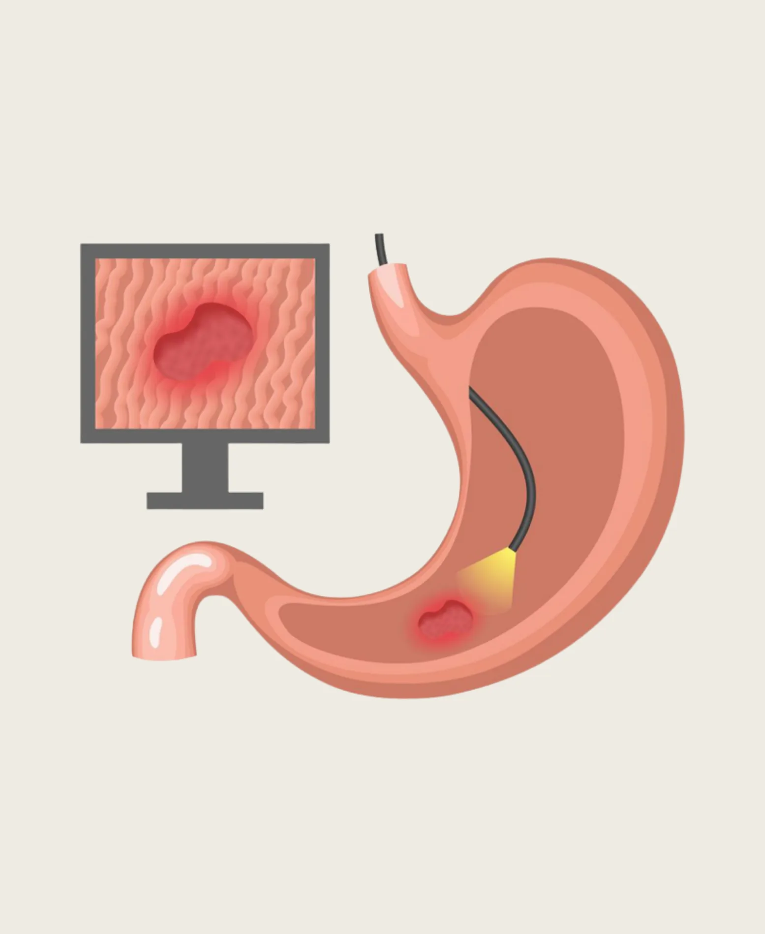
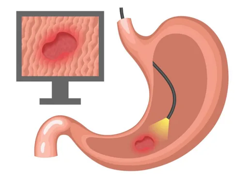
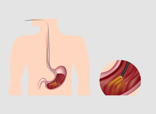
Upper GI endoscopy - also called OGD scopy, gastroscopy, or simply "camera test" - is the direct visual examination of the oesophagus, stomach, and the first part of the small intestine (duodenum) using a flexible fibre-optic or video endoscope: a thin, flexible, tube-shaped instrument (approximately 9 mm in diameter) with a high-definition camera at its tip.
The endoscope transmits real-time, magnified images to a high-definition monitor - allowing the endoscopist to assess the mucosal surface of the entire upper GI tract in detail, identifying abnormalities such as ulcers, erosions, polyps, masses, varices, Barrett's oesophagus, hiatal hernia, gastritis, and oesophagitis. Working channels within the endoscope allow the introduction of biopsy forceps, injection needles, electrocoagulation probes, haemoclips, and other therapeutic accessories.
Indications - when is upper GI endoscopy needed?
- Persistent heartburn, regurgitation, or acid reflux not adequately controlled by medication
- Dysphagia (difficulty swallowing - any cause)
- Epigastric pain lasting more than 4 weeks in patients over 45 (alarm symptom threshold)
- Haematemesis (blood in vomit) or melaena (black tarry stools)
- Suspected peptic ulcer disease - diagnosis and H. pylori biopsy
- Monitoring of Barrett's oesophagus
Treatment Approach
- Investigation of unexplained iron-deficiency anaemia
- Unexplained weight loss with upper GI symptoms
- Surveillance after gastric cancer surgery
- Pre-operative assessment before bariatric (weight loss) surgery
Therapeutic applications:
- Biopsy: Multiple biopsies taken from any suspicious mucosal area - sent for histopathological examination and H. pylori testing (rapid urease test - CLO test)
- Haemostasis: Control of acute upper GI haemorrhage from peptic ulcers - injection of adrenaline, thermal coagulation (heater probe, APC), or mechanical haemoclip placement
- Polypectomy: Removal of gastric or duodenal polyps using snare, cold biopsy forceps, or endoscopic mucosal resection (EMR)
- Oesophageal balloon dilation: Dilation of oesophageal strictures (peptic, post-surgical) or achalasia (pneumatic balloon dilation)
- PEG placement: Percutaneous Endoscopic Gastrostomy - insertion of a feeding tube directly into the stomach through the abdominal wall, under endoscopic guidance, for patients unable to swallow
Preparation:
Patients fast for a minimum of 6 hours before upper GI endoscopy (solids) and 2 hours (clear fluids). Regular medications should be discussed with Dr. Patel - antacids and anti-reflux medication may be continued; blood thinners may need temporary cessation.
Anaesthesia:
Upper GI endoscopy is performed under topical throat spray alone (for cooperative patients tolerating the gag reflex) or, more commonly, under conscious sedation - IV midazolam with or without fentanyl - which provides anxiolysis, reduces the gag reflex, and creates a degree of amnesia for the procedure. Most patients have no memory of the procedure and are comfortable throughout.
Duration: 10-20 minutes for diagnostic OGD. Therapeutic procedures (haemostasis, polypectomy) take longer.