01
Acute Appendicitis
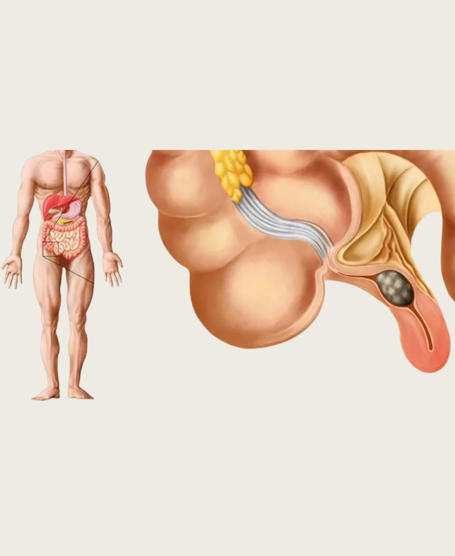
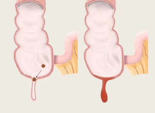
Acute appendicitis is inflammation of the appendix requiring urgent surgical removal. It is the classic cause of acute right lower abdominal pain in patients of all ages and remains one of the most important surgical diagnoses to make - or exclude - in anyone presenting with abdominal pain.
The clinical progression of appendicitis follows a predictable pattern:
Stage 1 - Early (0-12 hours): The appendix is distended and inflamed. Pain begins around the navel (periumbilical) - vague, crampy, and poorly localised. Nausea, loss of appetite, and sometimes a single episode of vomiting occur. Low-grade fever (37.5-38.5C). The patient is uncomfortable but not acutely unwell.
Stage 2 - Classical (12-48 hours): As inflammation spreads to the peritoneal surface (the lining of the abdominal wall), pain shifts to the right lower quadrant - specifically to McBurney's point (one-third of the way from the anterior superior iliac spine to the navel). The pain becomes localised, constant, and worsens with movement, coughing, or deep breathing. Rebound tenderness and guarding (involuntary tensing of abdominal muscles) indicate peritoneal irritation. Fever rises (38-39C). White cell count is elevated.
Stage 3 - Complicated (beyond 48-72 hours if untreated): The appendix wall necroses and perforates (bursts), releasing infected content into the peritoneal cavity. This causes generalised peritonitis - diffuse abdominal pain, board-like rigidity, high fever, and sepsis. This is a life-threatening emergency requiring immediate surgery and intensive post-operative care.
Diagnosis:
Clinical assessment combined with blood tests (elevated white cell count and CRP), urinalysis (to exclude urinary tract infection mimicking appendicitis), and imaging:
- Ultrasound abdomen: First-line imaging - can visualise an inflamed appendix in approximately 70-80% of cases (limited by overlying bowel gas and patient body habitus)
- CT abdomen and pelvis (with contrast): Gold standard - over 95% sensitivity and specificity for appendicitis, and also identifies alternative diagnoses when appendicitis is absent
- MRI abdomen: Preferred in pregnant women to avoid radiation exposure
Surgical treatment - Laparoscopic Appendectomy:
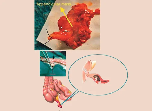
Laparoscopic appendectomy is the gold-standard treatment for acute appendicitis. Under general anaesthesia, three small incisions (one at the umbilicus, one in the lower abdomen, and one in the left lower quadrant) allow a camera and fine instruments into the abdomen. The appendix is identified, the mesoappendix (its blood supply) is divided, and the base of the appendix is secured with endoloops or a surgical stapler before the appendix is divided and removed. The procedure takes 20-40 minutes for uncomplicated appendicitis.
Advantages of laparoscopic appendectomy over open surgery:
- Smaller incisions - significantly less post-operative pain
- Allows thorough inspection of the entire abdominal cavity - important when the diagnosis is uncertain (allows alternative diagnoses to be identified and sometimes treated simultaneously)
- Lower wound infection rate - particularly important in females of reproductive age where the appendix incision can be near the fallopian tube
- Faster recovery - most patients are eating and mobile within hours of surgery
- Earlier hospital discharge - typically the next morning after a straightforward laparoscopic appendectomy
Recovery:
Hospital stay: 1-2 nights for uncomplicated appendicitis, 3-7 nights for perforated appendicitis. Return to desk work: 5-7 days uncomplicated; 2-3 weeks perforated. Full activity including sport: 3-4 weeks.